About your X-ray or scan in the radiology department
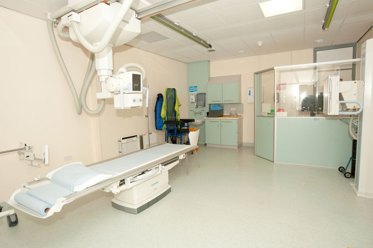
Going to have a scan or an X-ray can be daunting if you have never experienced one before. But there is nothing to be alarmed about as there are lots of people on hand to support you.
When you receive your appointment you may be given some instructions to follow such as having nothing to eat for 4-6 hours before the X-ray or drinking plenty of water so that you have a full bladder before the scan.
It is important that you follow these instructions and if they are not clear or you are unsure about any part of the procedure don't be afraid to ask questions.
Radiation safety
Your safety is very important as is the image quality of every X-ray.
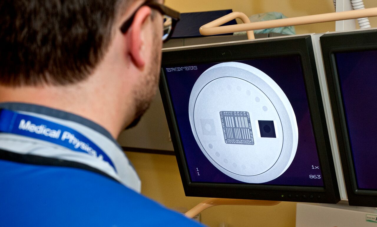
A medical physicist will check the image quality of an X-ray unit using a test tool.
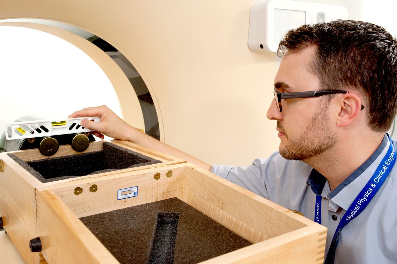
A medical physicist makes sure a test tool is level before testing a CT scanner.
Diagnostic imaging
You may be referred to the diagnostic imaging department for an investigation which will produce images of the relevant part of your body that will help in the identification, evaluation and monitoring of any disease processes or injury. The range of imaging investigations that may be done include:
Plain radiography
A radiograph is an image of the internal structures of the body and is produced by exposure to radiation (X-rays) with the image being recorded in digital form and displayed on a computer screen.
Fluoroscopy
Fluoroscopy is a procedure which uses radiation to produce a real-time image of parts of the body, where anatomy and function/movement can be assessed. In association with this study a contrast agent is used to outline parts of the body to be imaged, which would otherwise not show up well on the images.
Angiography
Angiography is a procedure where X-rays are used to investigate and image blood vessels and blood supply to body organs after injecting an iodine-based contrast agent into the vascular system (usually the femoral artery) via a catheter.
Computed Tomography Scanning
Computed tomography scanning provides cross-sectional images of the body using a beam of X-rays which rotates around the patient, and an array of sensors on the other side of the patient that detects the amount of radiation passing through the patient. This information is used to produce images that are displayed on a computer screen.
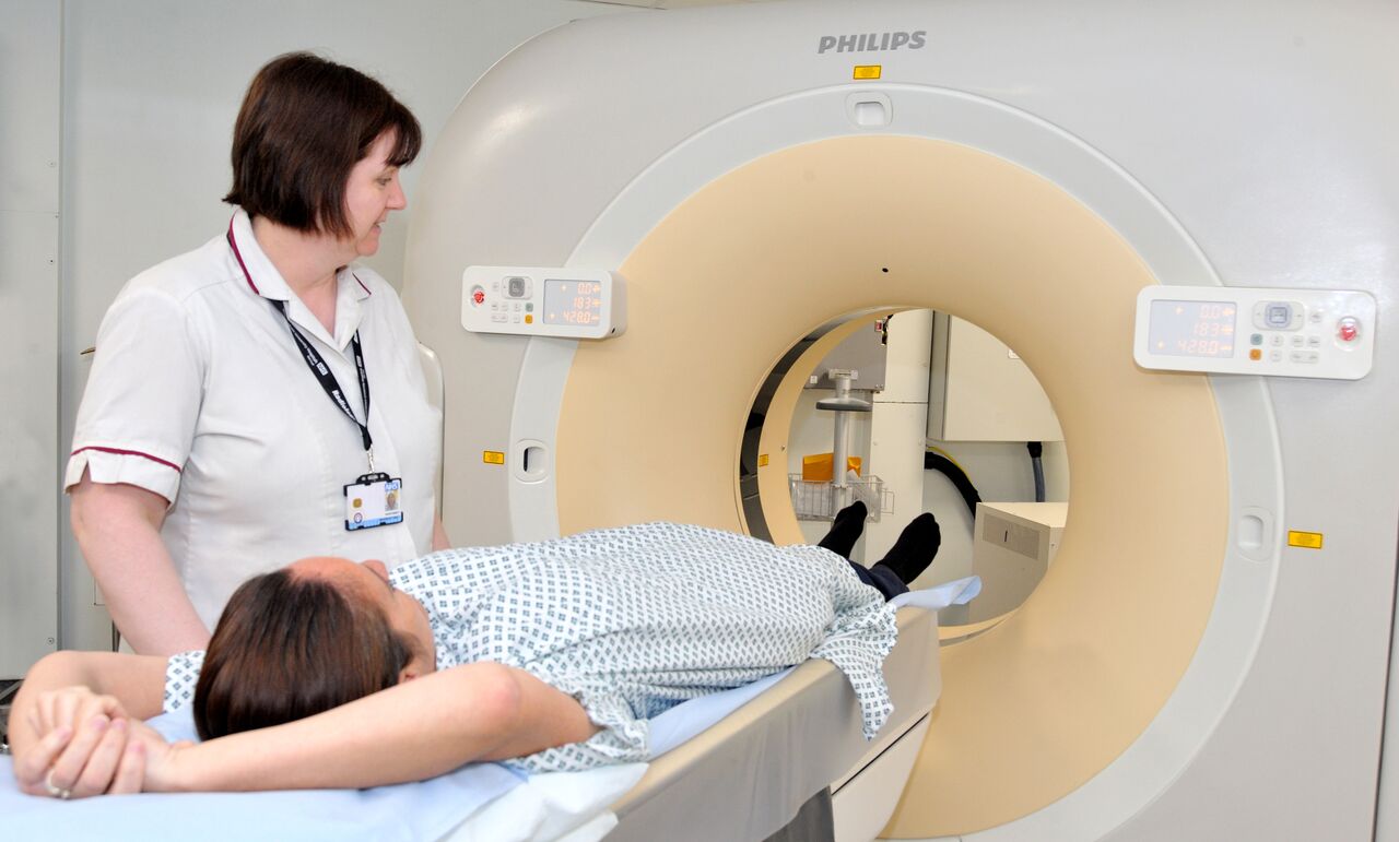
A radiographer will position the patient before a CT scan.
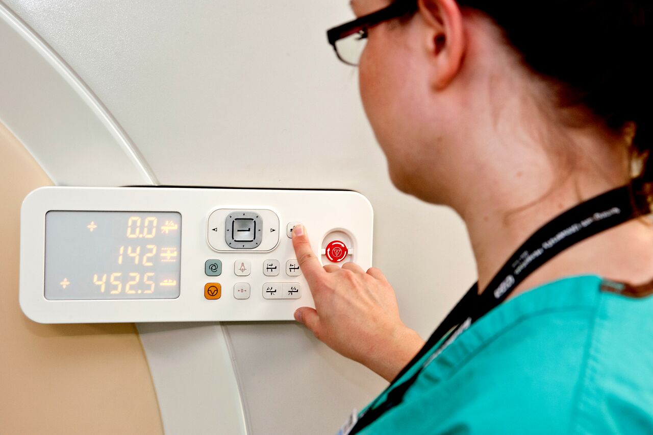
The radiographer will position the couch.
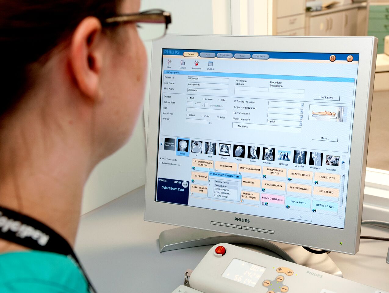
The type of scan is selected.
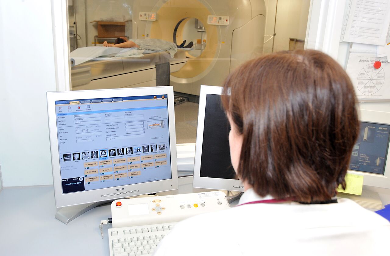
The radiographer begins the process of the scan from a computer.
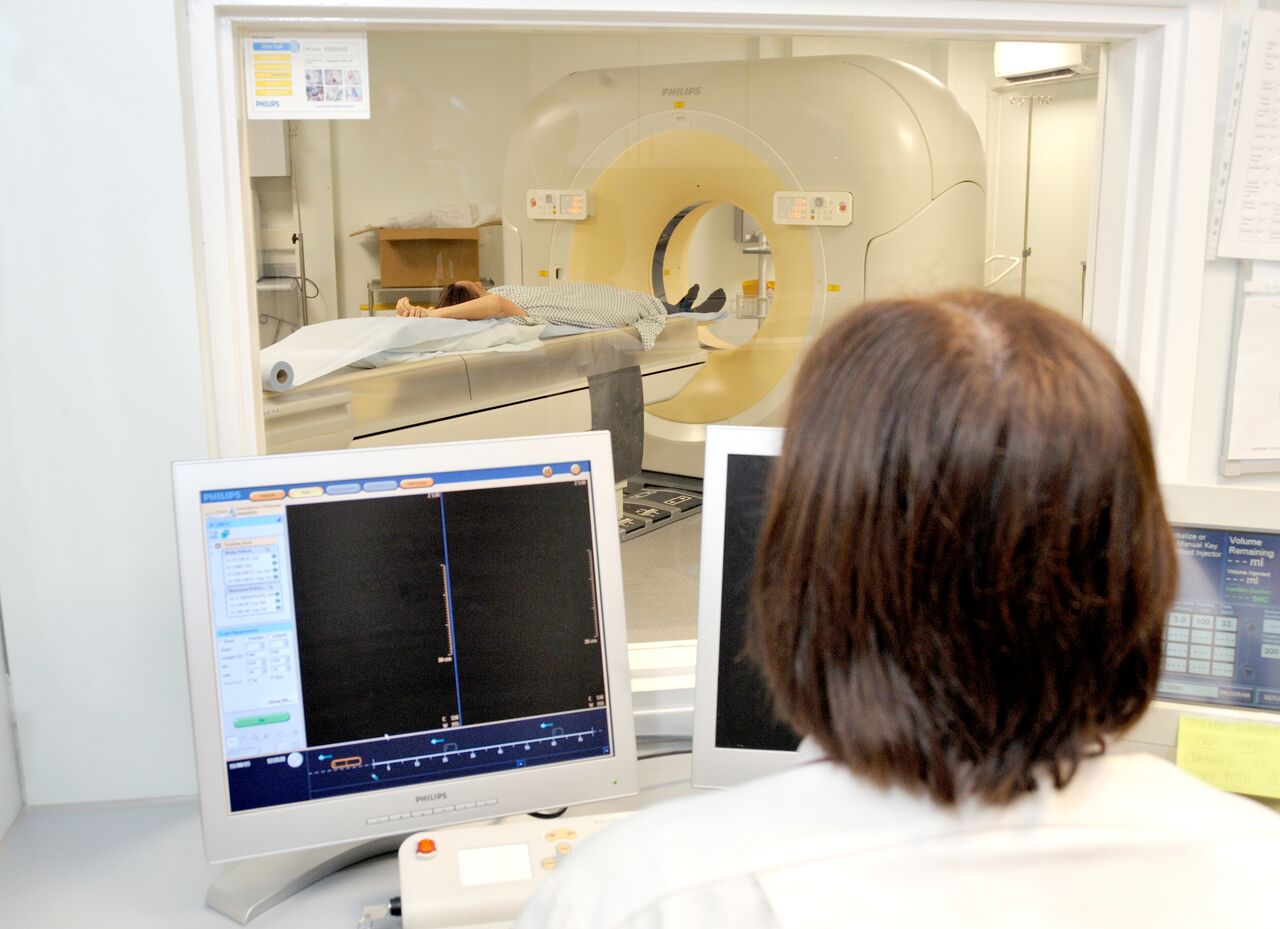
The radiographer observes the scan taking place from behind a screen.
Magnetic Resonance Imaging
Magnetic resonance imaging provides cross-sectional images of the body using powerful magnetic fields and radiofrequency pulses. The MRI machine produces a magnetic field, sends radio waves through the body, and then measures the signal response. The signals produced from the body by this process are used to generate images that are viewed on computer monitor.
Ultrasound
Ultrasound uses high frequency sound waves which are directed into the body and the sound is reflected as echoes from the internal organs. The echoes are detected and recorded to produce a real-time image of part of the body being investigated.
Nuclear medicine
Nuclear medicine uses radiopharmaceuticals, or radioactive drugs to examine how the body and organs function. Depending on the part of the body being imaged the radiopharmaceuticals are either introduced into the vascular system, swallowed or inhaled and accumulate in the relevant part of the body where it gives off a small amount of radiation as gamma rays. A gamma camera detects this radiation and the images produced give information on the structure and function of the area being investigated.
Interventional Radiology
This refers to a range of techniques which rely on the use of an imaging procedure to guide treatment to a specific area of the body. Most interventional treatments are minimally invasive starting with passing a needle through the skin to the area requiring treatment. This can be used to treat a range of conditions such as narrowing of arteries or removal of gall stones.
Images on this page are provided courtesy of Medical Physics, Nottingham University Hospitals NHS Trust